
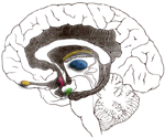
BRAIN ANATOMY
|
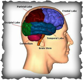
Brainstem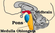
This is the lower extension of the brain which connects to the spinal cord. Located in the brainstem are the neurological functions that are necessary for survival (breathing, blood pressure, digestion, heart rate) and for arousal (being awake and alert).
The brainstem is connected to most of the cranial nerves and acts as a pathway for all the fiber tracts passing up and down from the highest part of the brain down to the spinal cord and peripheral nerves. |
Cerebellum

This is the portion of the brain that helps coordinate movement (balance and muscle coordination). It is located at the back of the brain. Damage to the cerebellum may result in ataxia which is a problem of muscle coordination, making it difficult for a person to perform everyday tasks such as the ability to walk, talk, eat and other self care tasks.
Frontal Lobe

This is the part of the brain which involves speech, thought, memory, organising, planning, problem solving, selective attention, personality and a variety of “higher cognitive functions” including behaviour and emotions. A deep groove or indentation called the central sulcus divides the parietal and frontal lobe. The frontal lobe is located at the front part of the brain. The anterior (front) portion of the frontal lobe in called the prefrontal cortex. The posterior (back) of the frontal lobe is divided into two sections, the premotor and the motor area. The prefrontal cortex is a very important area for the “higher cognitive functions” and the determination of the personality. The motor area consists of nerve cells that produce movement and the premotor area serve to modify movement. These muscular activities can be both voluntary and involuntary.
Occipital Lobe
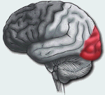
This region of the brain processes visual information and is located at the back of the brain. It is mainly responsible for visual reception however it also contains association areas that help in the visual recognition of shapes and colours. Any damage to the occipital lobe can cause visual deficits.
Parietal Lobe

They are superior to the occipital lobe and posterior to the central sulcus and frontal lobes. Their function is the perception of general sensation like touch, smell, temperature and conscious association. There are two parts of the parietal lobe, a right side and a left side. Damage to the right side can cause visuospatial deficits (patients may have difficulty finding their way around new or even familiar places). Damage to the left side may disrupt a patient’s ability to understand spoken and/or written language.
The parietal lobes contain the primary sensory cortex which controls sensation such as pressure and touch. Behind this is a large association area that controls fine sensation such as size, weight, shape and judgement of texture.
The parietal lobes contain the primary sensory cortex which controls sensation such as pressure and touch. Behind this is a large association area that controls fine sensation such as size, weight, shape and judgement of texture.
Temporal Lobe
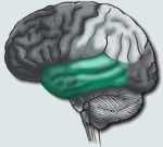
This region of the brain play an important role in organizing sensory input, auditory perception, language and speech production, as well as memory association and formation. There are two temporal lobes, one on each side of the brain. They allow a person to tell the difference between sounds and smells. The right lobe is mainly involved in visual memory e.g. memory for pictures and faces, whereas the left lobe is mainly involved in verbal memory e.g. memory for words and names.
Brain STRUCTURE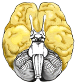
Cerebral Cortex
|
Function
This is composed of grey matter (nervous tissues containing neuronal cell bodies) and is the outermost layer of the cerebral hemisphere. Both hemispheres can analyze sensory data, learn new information, form thoughts, perform memory functions and make decisions.
|
Signs and symptomsNo signs or symptoms
|
Left Hemisphere
|
The left hemisphere is involved with the analysis sequential data such as systematic, logical interpretation of information. It’s also involved with the interpretation of symbolic information such as language, mathematics, abstraction and reasoning.
|
No signs or symptoms.
|
Right Hemisphere
|
The right hemisphere is involved with holistic functioning (relating to holism e.g. processing multi-sensory inputs). Additionally it is involved with one’s visual spatial skills. Dancing and gymnastics are examples of holistic functioning coordinated by the right hemisphere. Memory is stored in the right hemisphere in auditory, visual and spatial modalities.
|
No signs or symptoms.
|
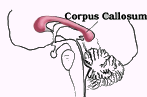
Corpus Callosum
|
The corpus callosum (a thick band of nerve fibers which divides the cerebellum into the two hemispheres) connects the right and left hemisphere to allow for communication between them. It transfers motor, sensory, and cognitive information.
|
Damage to this section of the brain may result in "Split Brain" syndrome.
|
Lymbic Symtem
|
The limbic system is involved with the Olfactory pathways (relating to the sense of smell). It includes:
|
Damage to the limbic system could cause loss of the sense of smell, agitation, loss of control of emotion and loss of recent memory.
|

Basal Ganglia
|
The basal ganglia are subcortical (beneath the cortex) gray matter nuclei. It’s involved with the initiation and direction of voluntary movement, balance and postural reflexes.
|
Damage to the basal ganglia may result in movement disorder: chorea, tremors at rest and initiation of movement. The sufferer will have difficulty initiating movement and have abnormal increase in muscle tone. Damage to this section of the brain causes certain nerve cells to die therefore causing the symptoms of Parkinson’s to appear. These could include tremor, rigidity and slowness of movement thus in effect cause tiredness, pain, depression and constipation. This can have an extremely negative impact on the sufferer’s lives.
|
Brain Anatomy and FunctionsView this incredible 3D animation showing the anatomy and functions of the brain using colour coded ares.
|
|
This page contains definitions and content belonging to other sources.
|
Copyright 2011-2012 Neurosurgeons.weebly.Com, Inc. All Rights Reserved.
|
Milad Rouf
|
